In Vitro Human Heart Microenvironments: Utilizing Cardiac Spheroids to Study Heart Disease
About this project
Project information
Exploring in-vitro human heart microenvironments using cardiac spheroids provides a powerful approach to study cardiovascular disease mechanisms and testing potential therapies. Cardiac spheroids are 3D cell culture models that mimic the architecture, cell composition, and microenvironment of human heart tissue. They are typically created from different cell types found in the heart, such as cardiomyocytes, fibroblasts, and endothelial cells which are cultured to form multicellular aggregates that replicate features of the heart, such as the myocardium's structure and function, thereby creating a model of cardiac tissue. This allows the study of complex cellular interactions that are pivotal in disease processes like fibrosis or hypertrophy. These spheroids are being used in high-impact functional studies, such as assessing contractility, measuring calcium flux, and evaluating metabolic activity. Cardiac spheroids are a versatile platform for cardiovascular research, enabling various applications such as molecular techniques, gene expression analysis, and protein expression studies. These spheroids facilitate immunostaining to visualize cellular interactions and conduct cytotoxicity assays to assess compound effects on cell viability. High-impact functional studies include evaluating contractility, measuring calcium flux, and investigating metabolic activity.
Research will focus on how both pathological exogenous and endogenous factors contribute to the induction of pathophysiological changes in cardiac spheroids. The research projects aim to uncover the mechanisms through which these factors disrupt normal cardiac function.
For more information about our research or collaboration opportunities, please contact geena.paramel@oru.se.
CS_Geena Paramel Video: Cultured human cardiac spheroid composed of cardiomyocytes, cardiac fibroblasts, and coronary endothelial cells.
Human cardiomyocytes in culture video: Human cardiomyocytes cultured in vitro for research purposes.

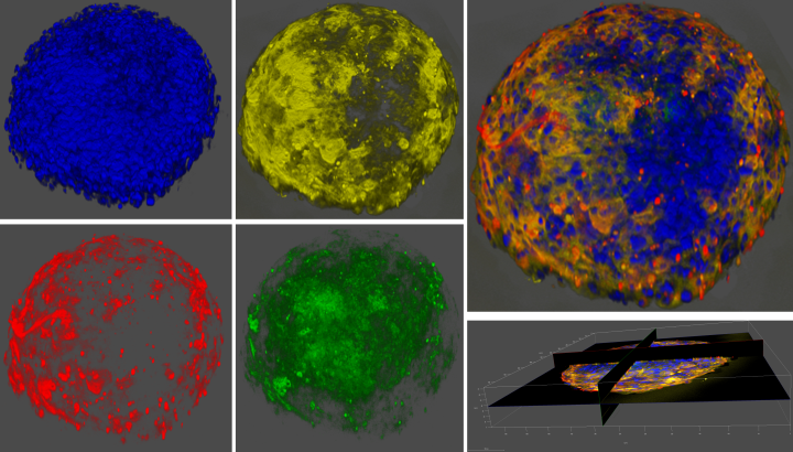
3D confocal microscopy images of human cardiac spheroid representing localization of cardiomyocytes (yellow), cardiac fibroblasts (red), coronary endothelial cells (green) and cell nucleus (blue).

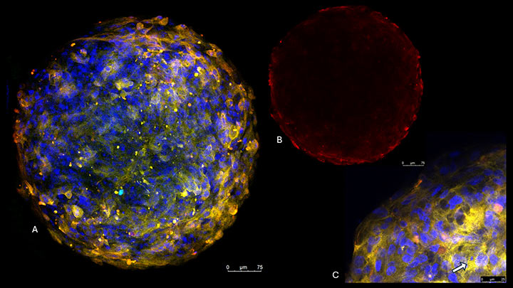
Fluorescent confocal microscopy images of human cardiac spheroids consisting of cardiomyocytes, cardiac fibroblasts, and coronary endothelial cells (A). Peripheral view of cardiac spheroids (Vimentin positive cells) (B). Cardiomyocytes staining showing Z-disc of sarcomeres (C).
